The goal of the DanMAX beamline is to do experiments on real material studied under realistic conditions at realistic time scales at multiple length scales. This is done by building three instruments at the beamline: One to study the atomic structure using PXRD in situ and in operando, one for studying the atomic structure in very high resolution, and one to study the microstructure using full field tomographic imaging. You can read more about the science at DanMAX in the Conceptual Design Report, here (PDF: 5 MB).
You can find all the publications from DanMAX here or on our Google Scholar site.
Experimental Techniques
Materials are extremely important in all aspects of our live and modern society would look very different without e.g. steel, advanced ceramics and doped semiconductors. Materials science and engineering is an interdisciplinary field focused on characterizing the structure, properties, performance, and processing of materials.
To understand and explain the properties of a material it is often essential to know the structure of the material. It is often necessary to know to study the structure at different length scales ranging from atomic detail to microstructure to get a complete understanding of the properties.
The structure at the atomic level is the arrangement of the individual atoms. An example of a structure-property relationship is graphite and diamond, which are both made up of carbon atoms. The microscopic structure concerns e.g. the grain and pore structure of the material. The microscopic structure governs e.g. the hardness of metals and the water permeability of chalk. Another example is chocolate where the right polymorph, i.e. crystal structure, gives nice shiny surfaces and the right ‘snap’ while the wrong crystal structure give dull looking and an undesirable texture.
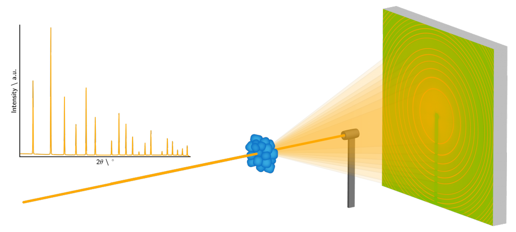
Powder X-ray diffraction
Powder X-ray diffraction (PXRD) is one of the most powerful ways to study the atomic structure of microcrystalline materials. Even though the experiment itself is simple the diffraction data contains a wealth of information. From PXRD data it is possible to get information about: Lattice parameters, symmetry, atomic coordinates, occupancies of the atomic positions, atomic vibration, quantitative phase analysis of crystalline mixtures, texture, domain size, micro and macro strain, defects, and even the local atomic structure.
Due to the simplicity of the experiment it is possible to use a wide range of sample environments to study materials under realistic working conditions. This combined with the immense intensity and low divergence of the MAX IV source allows for more complex materials to be studied.
The powder X-ray diffraction instrument at DanMAX is very versatile and can be used for studying e.g.:
- Formation and growth of nanocrystals and their structure under ambient and non-ambient conditions.
- The structure of battery materials in charging and discharging batteries.
- Identification of new hydrogen storage materials and other gas storage materials.
- Structural studies of functional magnetic materials.
- Materials performance and structure under load and processing in various environments.
- Structure and chemistry under high pressure.
- Bioinspired chemistry and biomineralization.
- Pharmaceutical science – control over polymorphs and structure solution.
- The structure and chemistry of catalysts under working conditions.
- … and much more…
Full Field Tomographic Imaging
Full-field tomographic imaging is a highly effective technique for examining the three-dimensional structure of materials at high resolution. This method leverages the power of X-ray phase contrast to reveal intricate details that are otherwise invisible with traditional absorption-based imaging. X-ray phase contrast can distinguish between different materials with similar densities, making it invaluable for studying biological specimens, polymers, and composite materials.
The simplicity of the full-field tomographic imaging experiment allows for diverse sample environments, enabling the study of materials under various conditions. With the intense X-ray beam from MAX IV, this technique can probe more complex and heterogeneous materials. This capability extends the range of potential applications, from characterizing internal defects in hard materials to understanding the internal structure of soft tissues and the flow of liquids through materials.

The X-ray imaging instrument at DanMAX is very versatile and can be used for studying e.g.:
- The microstrucutre structure of battery materials in charging and discharging batteries.
- The structure of biological and medical biopsy samples.
- Materials performance and structure under load and processing in various environments.
- Structural studies of functional engineered materials.
- Flow of media through a material.
- 3D structure of fossils.
- … and much more…