Follicular tumours in the thyroid can be difficult to diagnose as the entire follicle capsule needs to be sliced and inspected in order to detect ruptures. The current protocol involves cytology and histology, but these have limitations. Researchers from Uppsala University (UU) and Lund University (LU) are investigating the potential use of synchrotron-based virtual histology for 3D inspection of the follicle capsule at MAX IV.
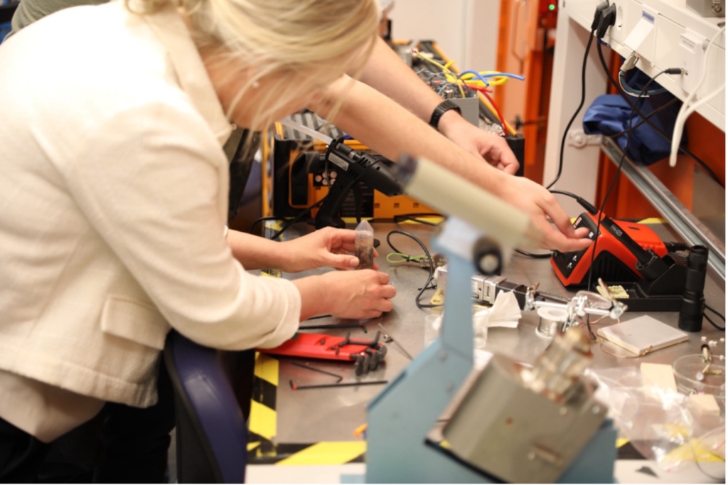
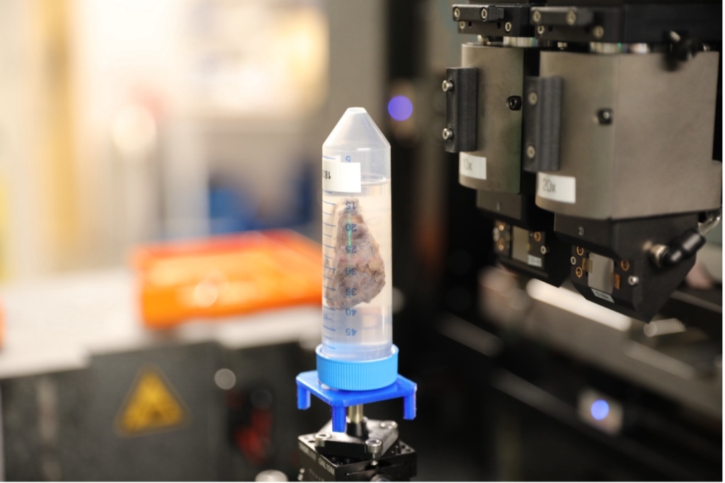
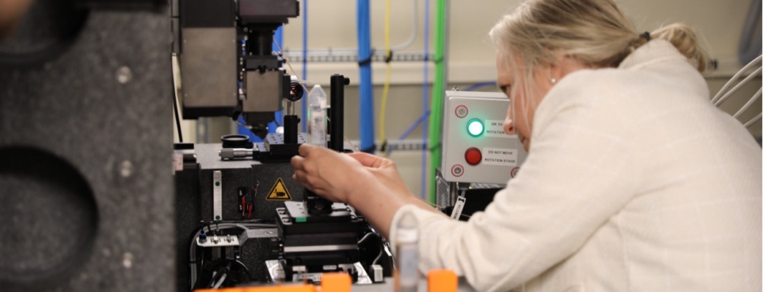
Thyroid tumours can be either benign follicular adenoma or malignant follicular carcinoma. The ability to assess the difference is crucial. Although cytological analysis can effectively distinguish between benign and malignant, it is unable to detect key diagnostic indicators of follicular carcinoma such as capsular breach or vascular invasion. In addition, further detailed histopathological analysis following diagnostic surgery is often required, which can be meticulous and time-consuming due to the number of thin slices necessary to correctly identify whether malignant indicators are present. Lund University Associate Professor Martin Bech and resident Physician Matilda Annebäck and Chief Physician Olov Norlén from Uppsala University conducted a pilot study at MAX IV’s DanMAX beamline to determine the applicability of a new and improved assessment method for thyroid tumours.
Synchrotron radiation-based micro-tomography (SRµCT) is an imaging technique that enables 3D mapping of internal structures of materials. At DanMAX, the field of view is 1.2 x 1.2 mm, allowing analysis of the thyroid lobes and, with exceptional spatial resolution, enabling detailed 3D visualization of the thyroid tissue.
Learn more about open access for academic research at MAX IV.
Are you interested in industrial research and collaboration possibilities? Read more here.
The goal is that SRµCT will detect those diagnostic features that are missed or difficult to spot with current available methods. Another advantage of SRµCT is that it is non-destructive to the sample, and thus will allow for subsequent histological analysis for comparative analysis between the two techniques

This will be the first study using SRµCT for follicular thyroid tumours, although it has been conducted elsewhere in other tissues from the lung, heart and brain. The challenge in the current experiment was the sample size in relation to the beam size. As a proof-of-concept experiment, it was deemed successful.
MAX IV is an ideal location for clinical experiments in Sweden as ethical approval often has stipulations about transport of samples outside of the country. The study samples were used for diagnostic purposes thus it was not difficult to obtain the ethics approval.
The collaboration between experts in the thyroid field (Department of Surgical Sciences; Endocrine Surgery, UU), and experts in SRµCT (X-Ray Phase Contrast Group, LU), arose after Martin Bech presented at Uppsala about the possibilities at MAX IV, with Olof Norlén in attendance at the talk.
This is a great example of how many studies are conducted at MAX IV and illustrates the need for mixed expertise in order to conduct these experiments, as the researchers from UU have the clinical expertise but were new users at MAX IV. As both parties are part of academic institutions, they were able to apply for free access to MAX IV through the peer-reviewed process which negates the need to apply for separate experimental funding.
SRµCT experiments generate substantial data, which is currently being analysed by the pathologist at UU and compared to their histological findings. More detailed data analysis will be another collaboration between the two research groups with the ultimate goal to publish the findings. This technique is of great interest to pathologists and has further implications both in the research and clinical fields.

The imaging end station at DanMAX beamline opened for users in 2024 and brings the total to 4 beamlines at MAX IV with imaging capabilities. A unique feature of DanMAX is the X-ray Powder Diffraction end station which can be used for studying atomic structure, especially of microcrystalline materials.
With two end stations, DanMAX has applications in many fields such as life science, the pharmaceutical industry, cultural heritage, material science and many more. The availability of sample environments for varying heat, pressure, humidity etc., enables experimentation under realistic conditions and for both in situ and operando experiments.
Are you interested in using MAX IV through open access? Find out more here.
Are you in industry and want to utilise MAX IV? Find out more here.


