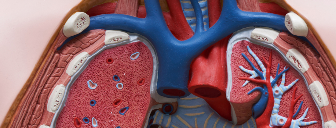
Researchers have imaged lung tissue affected by Idiopathic Pulmonary Fibrosis (IPF) with nanometre resolution. They managed to capture differences in the distribution of trace elements compared to a healthy lung. The result is a step towards better understanding the body at the nanoscale and managing this and other currently untreatable diseases.
The distribution of chemical elements in our cells can say a lot about their function and processes. To see this distribution, we need a method with a resolution that is high enough to see details inside cells. The method should also be sensitive to differences in chemical content. The structures inside the cell are on the nanometre scale, and Nano X-ray Fluorescence (Nano-XRF) offers a powerful imaging method that fulfils both criteria. The technique is relatively young and has been used in the research community for about 10–15 years.
“Nano-XRF is becoming more prominent as synchrotron facilities are advancing. Its growing use is linked to improvements in synchrotron technology, such as brighter beams and better focusing, that now allow nanometre-scale spatial resolution and higher sensitivity, enabling applications that were not possible before,” says Bryan Falcones, a postdoc at the Department for Lung Biology at Lund University and visiting research fellow at MAX IV.
IPF could be connected to the inhalation of different types of environmental pollution, with particles being inhaled and stuck in the lungs, where they irritate the cells. The researchers found metals accumulating around carbon particles in the IPF-affected tissue. They also found metal clusters of zinc, calcium, and colocalization of iron with sulphur. The explanation for the findings is, however, not yet clear, and the research project continues.
“At the moment, we are exploring larger cohorts of IPF-tissue donors to connect our findings with disease symptoms. We are also extending our scans to other chronic diseases, such as chronic obstructive pulmonary disease or COPD, as well as investigating the impact of cigarette smoke on airway epithelia. Finally, we are delving deeper into iron chemistry in lung diseases, including other X-ray techniques,” concludes Bryan Falcones.